Welcome to the Future of Eye Exams
One of the most important parts of a comprehensive eye exam is determining the health of the retina. The retina is made up of nerve cells and blood vessels that connect to the brain and body, and it gives your doctor an important window into your overall health. In fact, the blood vessels in the retina are the only place that a doctor can see blood vessels in the body without cutting the body open! As a result, eye doctors often detect diseases like diabetes, hypertension, and elevated cholesterol before other physicians.
For many years, the only way to get a good view of the retina was to dilate the eyes. This process involves the doctor placing special eye drops into your eyes to open up the iris (the color part of the eye). When the iris is open, the doctor uses a bright light and lenses to examine the nerves and blood vessels of the retina for signs of damage or disease.
Unfortunately, in spite of its benefits, dilation is not a pleasant experience for most patients. In fact, many patients refer to it as the most “annoying” part of the eye examination. People generally do not like having drops in their eyes (especially if they are sensitive to the preservatives that can make the eyes burn for several seconds afterward), they don’t like waiting 10-15 minutes for the drops to work, and they typically do not enjoy the bright lights from the doctor or their surroundings until the dilation wears off 3-4 hours after the examination.
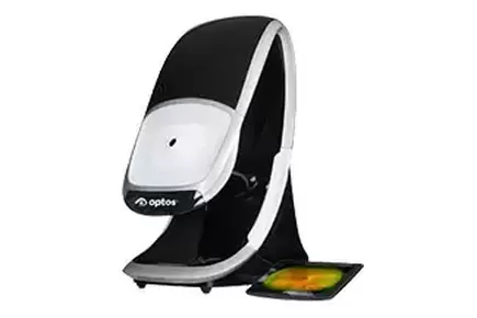
Thankfully, we now have an alternative to dilation at Atlantic Family Eye Care called the optomap. This high-tech camera takes a wide-field, high resolution, digital picture of the retina – without eye drops! It is fast, painless, and as comfortable as getting your picture taken. It is safe to use on the whole family, and the parents of young children in our practice really love it because their kids don’t have to get drops in their eyes!
In addition to being faster and more convenient than dilation, the optomap also allows the doctor to show each patient what the back of their eye looks like. At our practice, we review the pictures with the patient during the examination and explain what we are seeing in the nerves, blood vessels, and overall health of the retina. Instead of “annoying,” this is now one of the most anticipated and rewarding aspects of the examination for our doctor and patients alike because it gives them a snapshot of their overall eye health and provides an opportunity to discuss any questions or concerns that arise from any abnormal findings.
Another advantage that optomap retinal imaging has over traditional dilation is the ability to easily detect retinal changes over time. If a patient has an abnormal finding, like broken blood vessels from diabetes, in the back of the eye, it is easily detected by a skilled eye doctor with dilation or the optomap. But how does the doctor determine if it is changing or getting worse from year to year? With dilation, they have to compare their findings to previous chart notes and hope they are detailed enough to provide the most precise information; but, with the optomap, the doctor puts the pictures side-by-side on the computer screen and can easily tell if there are differences. Our patients at Atlantic Family Eye Care repeatedly tell us how much they appreciate seeing and knowing that we have the most accurate information possible for evaluating the health of their eyes.
While the optomap has been a great addition to our practice, it is not a replacement for dilation. There are several instances where dilation is more appropriate than the optomap. These include patients with extremely small pupils, droopy eyelids, dementia, certain physical disabilities, and emergency eye conditions.
At Atlantic Family Eye Care, we believe that dilation and the optomap are both important tools for evaluating the health of the retina, but one or the other may be a better option for you depending on your circumstances. So next time you visit our office, please ask us which one is right for you.